The muscle that allows for an outward rotation of the paw.
Supinator m.
The superficial vein of the antebrachium that is often used of venipuncture.
Cephalic v.
The bones of the antebrachium.
Radius and Ulna
This bone is absent or reduced in most cursorial animals, but provides boney attachment of the thoracic limb to the thorax in most biped, brachiators, manipulators, and diggers.
Clavicle.
The three layers of embryonic development (i.e. the 3 germ layers)
Mesoderm, Ectoderm, Endoderm
The flexors of the elbow joint.
Biceps brachii m. and brachialis m.
The nerve of the triceps brachii.
Radial n.
The grove where the anconeal process of the ulna sits during extension.
Olecranon fossa.
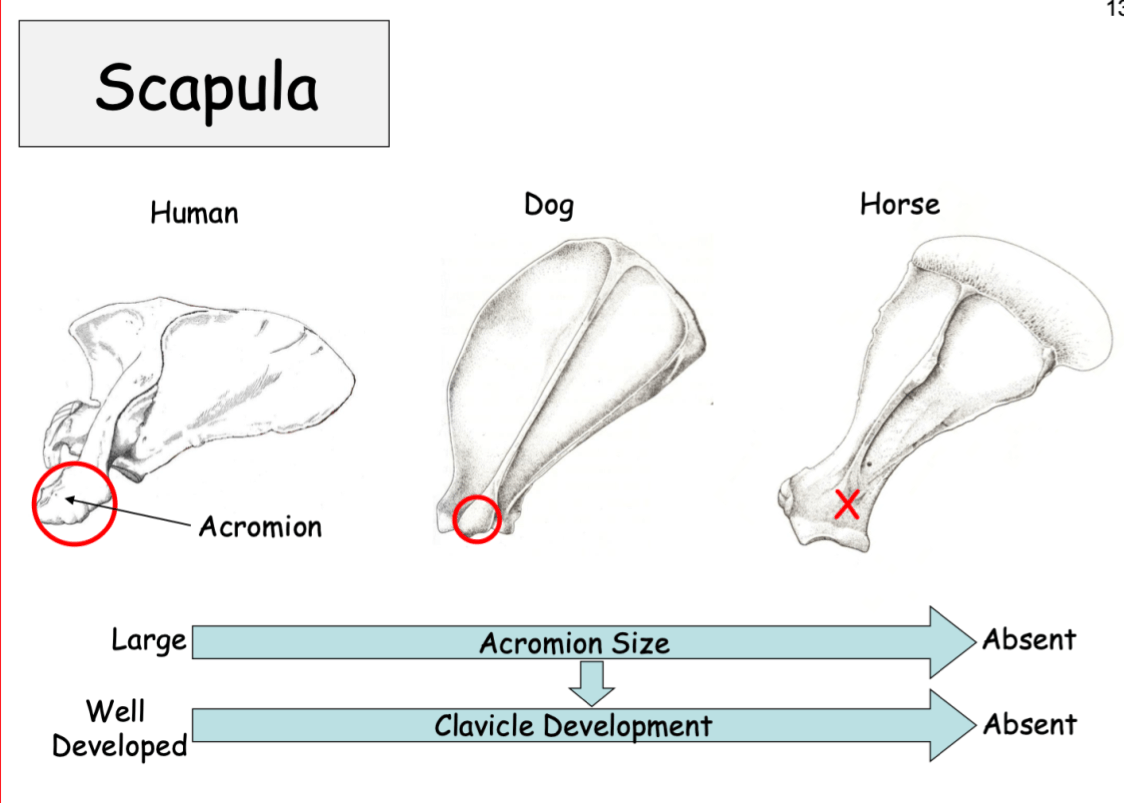
This boney prominence is reduced in dogs, and absent in horses, and its size is often correlated with the clavicle development.
Acromion
The solid ball of cells inside the zona pellucida as a result of cleavage, usually 16-64cells in size.
Morula
The muscles that comprise the "caudal or flexor group" of the shoulder.
Deltoideus m., Teres minor m., Teres major m.
The nerve that originates from the spinal segments C8 and T1, and innervates the Flexor carpi radialis m. and the Superficial digital flexor m.
Median n.
The equine insertion point for the Flexor carpi ulnaris.
Accessory carpal bone
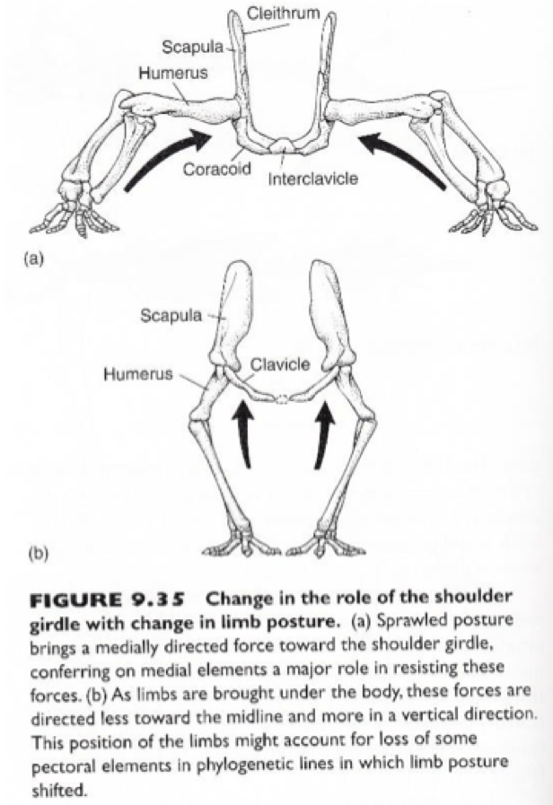
In the "original" animal design the limbs sat out laterally from the body (as seen in most lizards today) and this provided these animals with a lot of stability. Through evolution the limbs are now under the trunk of the animal, there are advantages and disadvantages, name one of each.
Advantages:
1) decreases the loads in the ventral girdle of the animal
2) increased protraction and retraction of the limb = faster
Disadvantages:
1) narrower base of support = less stable
2) reduced latero-flextion of the body
The aspect of the blastocyte where the cells in that area become larger and will eventually the area from which the embryo will develop.
Embryonic disc.
Serratus ventralis m.
Pectoralis profundus m.
The nerve of the shoulder "flexor" group.
Axillary n.
The type of joint of the thoracic girdle to the axial skeleton.
Synsarcosis: a junction of two or more bones by means of muscular attachment(s).
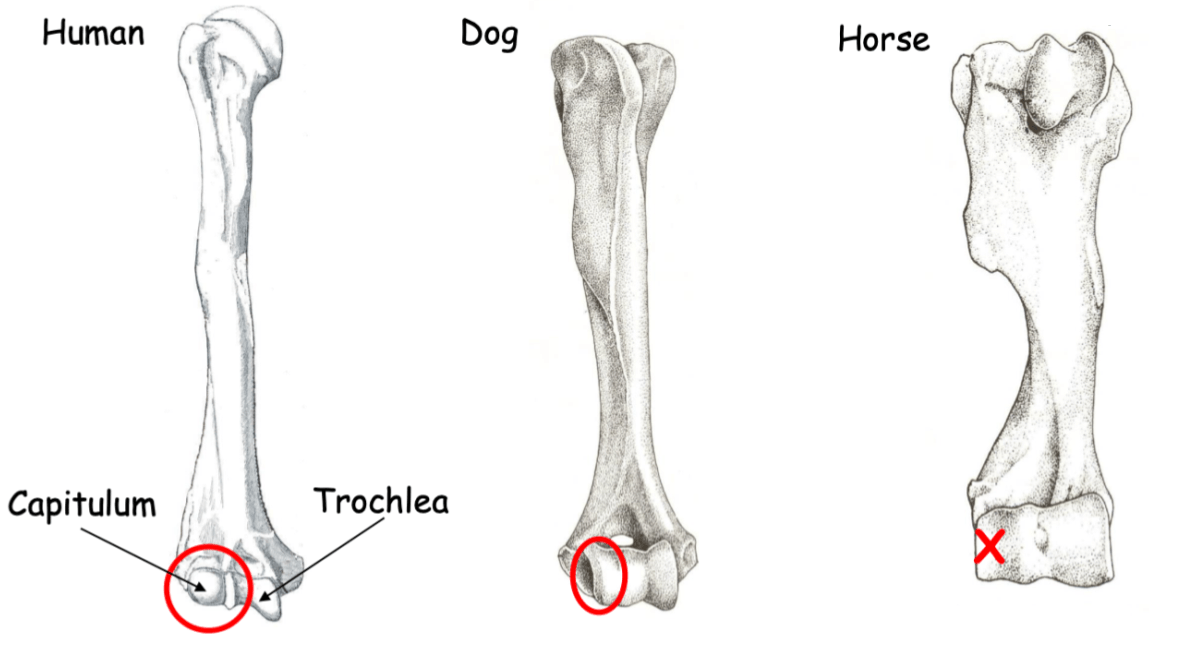
This boney prominence is absent in horses, is partially present in carnivore, and fully developed in manipulators. This boney prominence increased the range of movement of the forelimb and is on the distal end of the humerus.
Capitulum (it engages with the radius)
The cells around the blastoceol, cells which facilitate absorption of nutrients. These cells are not found on the embryonic disc in all animals expect primates.
Trophoblast cells.
(Greek: troph - nourish, blast - germ)
The muscle that draws the thoracic limb cranially, with an insertion on the wing of the atlas.
Omotransversarius m.
(Origin: distal scapular spine)
The nerve that is most commonly damaged in trauma to the proximal forelimb, especially in instances of fractures.
Radial n. b/c it wraps from the caudal aspect of the humerus to the cranial distal part of the humerus. Comes from the rotation of the limbs due to evolution.
The bones of the equine mid- or inter-carpal joint.
Proximal (medial to lateral): Radial carpal bone, intermediate carpal bone, ulnar carpal bone.
Distal (medial to lateral): 2nd carpal bone, 3rd carpal bone, 4th carpal bone.
(the 1st carpal bone is frequently isolated from the remainder of the skeleton, embedded in the palmar carpal ligament behind the second carpal)
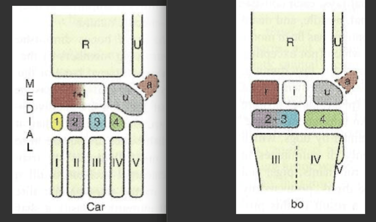
Compare and contrast the carpal bones of a bovine vs a carnivore.

The embryological cell layer making up the neural tube.
Ectoderm.